Epulis in Dogs: How Is It Diagnosed, and Does It Need To Be Treated?



vadimguzhva/iStock/Getty Images Plus via Getty Images
An epulis is a growth that can be found on the gums inside a dog’s mouth. Epuli in dogs are common and typically benign (noncancerous).
Recently, there was a name change for this condition and restructuring of how veterinarians group the different types of epulis in dogs’ mouths.
Previously, there were three types of epulis: fibromatous epulis, ossifying epulis, and acanthomatous epulis. You may hear new terms for these types of gum growths that we’ll discuss in more detail.
Let’s explore these growths inside the mouth and what it means for your dog if they have an epulis.
Key Takeaways
- An epulis is a growth on a dog’s gums.
- Epuli or epulides (plural for epulis) are generally considered noncancerous.
- There are three different types of epulis based on the type of cells that make up the mass.
- Treatment is based on how aggressive the epulis is.
What Is an Epulis in Dogs?
An epulis is a nonspecific term that can be used for any growth on the gums. Historically, epulis-type growths have been considered benign, or noncancerous.
An epulis can look like a pink growth on the gums above or around the teeth. They will often trap bacteria near the teeth, leading to more dental disease as plaque builds up.
The growths form when the periodontal cells of the gums divide and multiply abnormally.
Epulis can be put into three different categories based on the cell type of the growth. The reason for this distinction is that the three different types of periodontal growths behave differently and need different treatment.
These gum growths are classified as one of three types of periodontal tumor:
- Peripheral odontogenic fibroma (POdF): This is the most common type of epulis. This growth is made up of tough connective tissue. This growth was formerly known as a fibromatous epulis.
- Peripheral ossifying fibroma (POF): Like the common masses above, this type also includes some bone tissue inside the growth. It was previously called an ossifying epulis.
- Acanthomatous ameloblastoma: This is the rarest and most aggressive type of epulis. Ameloblastomas are locally invasive and grow into the underlying bone of the jaw, causing breakdown of bone, jaw swelling, and pain. This growth was previously called an acanthomatous epulis.
Epuli are generally not considered a medical emergency.
However, if your dog has a bleeding mass or lump on their gums and is not eating, or is otherwise acting sick and unlike themselves, they may need to be seen on a more urgent basis.
Symptoms of an Epulis in Dogs
If your dog has an epulis, you may notice:
- Visible mass on the gums
- Drooling
- Halitosis (bad breath)
- Bloody saliva
- Facial swelling
- Difficulty eating
- Chattering the jaw
Causes of an Epulis in Dogs
Epulis growths are caused by abnormal division of cells. This can come from genetics, overcrowding of the teeth, and chronic trauma or irritation of the gums.
Genetics can play a role in whether your dog gets an epulis.
Certain breeds are more likely to get epulis in their lives, including:
- Boxers
- Border Terriers
- Basset Hounds
- Shetland Sheepdogs
- Golden Retrievers
- Akitas
- American Cocker Spaniels
Male dogs are statistically more likely to get epulis than female dogs, and they are more common in middle aged and senior dogs.
Overcrowding of the teeth can lead to more friction from the teeth rubbing on the gums. This chronic irritation of the gums may predispose them to getting gum tumors.
In a similar fashion, dental disease can lead to chronic inflammation (swelling) of the gums and then tumors forming.
How Veterinarians Diagnose an Epulis in Dogs
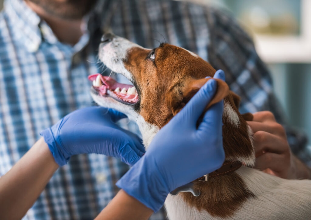
Epulis growths can usually be seen on a physical exam by a veterinarian.
Your vet will start with an oral exam. If a mass is found on the gums, they will likely recommend surgical removal and histopathology (biopsy) of the growth. Unfortunately, you cannot tell just from looking at a tumor if it is cancerous or not.
If your dog has swelling of the jaw, your vet may recommend X-rays of the face and/or dental radiographs.
Lab work may also be recommended to ensure organ function is good before sedation or anesthesia.
Treatment of an Epulis in Dogs
Treatment for an epulis starts with surgery. The growth will be removed and sent to a laboratory for histopathology (biopsy). This will help decide the next steps.
Treatment moving forward depends on which type of epulis is diagnosed.
If the epulis biopsy results come back as a POdF or POF, then removing the mass usually cures the condition.
Sometimes the neighboring tooth must be taken out as well to keep the mass from growing back.
If the epulis biopsy results come back as an acanthomatous ameloblastoma, then treatment is much more aggressive.
This rare form of epulis will invade and destroy the jawbone, leading to chronic pain and swelling. Surgery is often major and may involve removing part of the jaw.
Radiation therapy is also often done in dogs who had ameloblastoma to keep the mass from growing back.
Your dog will likely be prescribed pain medications, like carprofen and/or gabapentin post-operatively.
Your pup may also be put on antibiotics after surgery, depending on how big the surgery was and whether infected teeth were involved as well.
Antibiotics like clindamycin or Clavamox may be prescribed.
Recovery and Management of an Epulis in Dogs
Following removal of the epulis, your dog will likely need to be on a bland, canned food, such as Hill’s Digestive Care, or a softened food diet for the next 10–14 days while their gums are healing.
Avoid letting them chew on anything with a crunch that may irritate the gums during the recovery period.
After having an epulis removed, it typically takes 10–14 days for the gums to heal.
Your vet may recommend a recovery cone, such as this WZ PET cone, depending on the location of the epulis and whether any dental extractions were done during the procedure.
Prevention of an Epulis in Dogs
As genetics play such a large role in whether your dog may get an epulis, there is not a lot that pet parents can do to prevent them.
Regular dental care can reduce their risk. One of the contributing factors to an epulis forming is chronic irritation and inflammation of the gums.
The Veterinary Oral Health Council has published a list of products they recommend for routine dental care.
Using VOHC-recommended dental products, like OraVet chews or PlaqueOff, may reduce buildup of tartar in the mouth.
Routine dental cleanings can also help keep your dog’s mouth healthy.
Epulis in Dogs FAQs
Can an epulis in dogs go away on its own?
An epulis is a mass that will typically not go away without being surgically removed.
Is an epulis in dogs cancer?
An epulis is typically benign and not considered cancerous. However, the rare ameloblastoma type of epulis can be locally aggressive and may need to be treated in a similar fashion as cancerous oral growths: with aggressive surgery and radiation.
What does an epulis look like in a dog's mouth?
An epulis looks like a pink mass, lump, or bump on the gums.
Is an epulis painful for dogs?
While most epulis growths are not painful, an epulis can become painful if it invades the jawbone or if it is trapping plaque, leading to an infected tooth nearby.
Can an epulis come back after surgery?
An epulis can come back after surgery. Sometimes removing the nearby tooth may be recommended to reduce the risk of the epulis returning in the future.
Are certain dog breeds more likely to develop epulis?
Certain breeds are more likely to develop an epulis. Boxers, Border Terriers, Basset Hounds, Shetland Sheepdogs, Golden Retrievers, Akitas, and American Cocker Spaniels all have an increased risk of getting an epulis.
Should I be worried if I find a lump on my dog's gums?
If you find a lump on your dog’s gums, you should have it checked out by a veterinarian. They will likely recommend removing the mass and sending it off for biopsy to find out if it’s cancerous or not.
References
Peripheral Odontogenic Fibroma in Dogs: Vet Clinical Guide. International Veterinary Dentistry Institute. Published 2025. https://veterinarydentistry.net/peripheral-odontogenic-fibroma/


