Corneal Ulcers in Guinea Pigs: Everything a Pet Parent Needs To Know


monkeybusinessimages/iStock/Getty Images Plus via Getty Images
If your guinea pig is squinting, tearing, or suddenly has a cloudy eye, they may have a corneal ulcer—a painful scratch or injury to the eye’s surface that needs prompt veterinary care.
Fast treatment is important because even small ulcers can quickly worsen, become infected, and threaten your guinea pig’s vision.
Key Takeaways
- Corneal ulcers in guinea pigs are painful eye injuries that can quickly worsen without veterinary treatment and may lead to infection, vision loss, or eye rupture.
- Common signs of a corneal ulcer in guinea pigs include squinting, watery or cloudy eyes, redness, pawing at the eye, and decreased appetite or activity.
- Most guinea pig corneal ulcers heal with prompt veterinary care, proper eye medications, and a clean, low-dust habitat that helps protect the eye during recovery.
What Are Corneal Ulcers in Guinea Pigs?
A corneal ulcer in guinea pigs is a small defect or damage to the cornea, which is the surface of the eyeball or the dome-shaped outer layer covering the pupil and iris.
The defect can be as small as a tiny scratch, or it can be quite large to the point of perforation (a hole) where the inner contents of the eyeball can bulge out.
These defects in the cornea are very painful, and the pain will cause excessive tearing and squinting. There is also often redness around the eye. The eyeball can start to look cloudy, blue-white, or red because of the injury and inflammation (swelling).
Sometimes the scratch can be so small you will not see a change to the cornea, but if there is some redness, squinting, or excessive tearing, having your guinea pig checked out is very important because corneal ulcers can be common in guinea pigs.
An ulcer happens when the outer layer of the cornea is damaged. This typically happens from trauma, such as hay poking into the eye, rough bedding poking the eye, or a scratch from another animal.
Guinea pigs also produce less tears compared to other mammals, which can make them more at risk of corneal issues.
A corneal ulcer should be treated urgently. They are very painful and can easily become infected. Small ulcers that aren’t treated can also become deeper ulcers and cause a rupture of the eye or possible permanent vision loss.
Symptoms of Corneal Ulcers in Guinea Pigs
Symptoms of corneal ulcers in guinea pigs include:
-
Squinting, keeping one eye closed
-
Watery eye discharge
-
White, cloudy, blue, or gray discoloration of the eye
-
Pawing at the eye
-
Sensitivity to light or shying away from bright lights
-
Hiding, lethargy, less of an appetite
-
Thick yellow or green eye discharge
-
Redness around the eye or swelling
Causes of Corneal Ulcers in Guinea Pigs
The most common cause of corneal ulcers in guinea pigs is an eye injury.
This can happen from hay, improper bedding, toys, or another guinea pig scratching the eye. Decreased tear production can also make them more at risk of corneal damage.
Risk factors can include:
-
Hay poking the eye if too coarse or rough
-
Dusty or rough bedding causing eye irritation
-
Fighting with a cage mate
-
Underlying eyelid problems like an eyelid mass or the eyelid rolling inward
-
Cage accessories that are sharp, broken, or rough
-
Bacterial infection or upper respiratory infection
-
Hair or eyelashes that are poking into the eye
There is not a sex or age tendency for this problem. But poor environmental conditions can put guinea pigs at higher risk for corneal ulcers.
How Veterinarians Diagnose Corneal Ulcers in Guinea Pigs
Veterinarians diagnose corneal ulcers by looking at the eye and doing special tests.
Pet parents should not start any leftover eye medication from earlier visits unless directed by their veterinarian because it can sometimes make the condition worse.
Steroid eye drops will often worsen ulcers. And most eye drops only last about 30 days.
Using them past that time frame puts your pet at risk for infection due to bacterial growth in the bottle.
Your veterinarian may do:
-
Physical exam: To look at overall health, hydration, teeth, and any other abnormalities
-
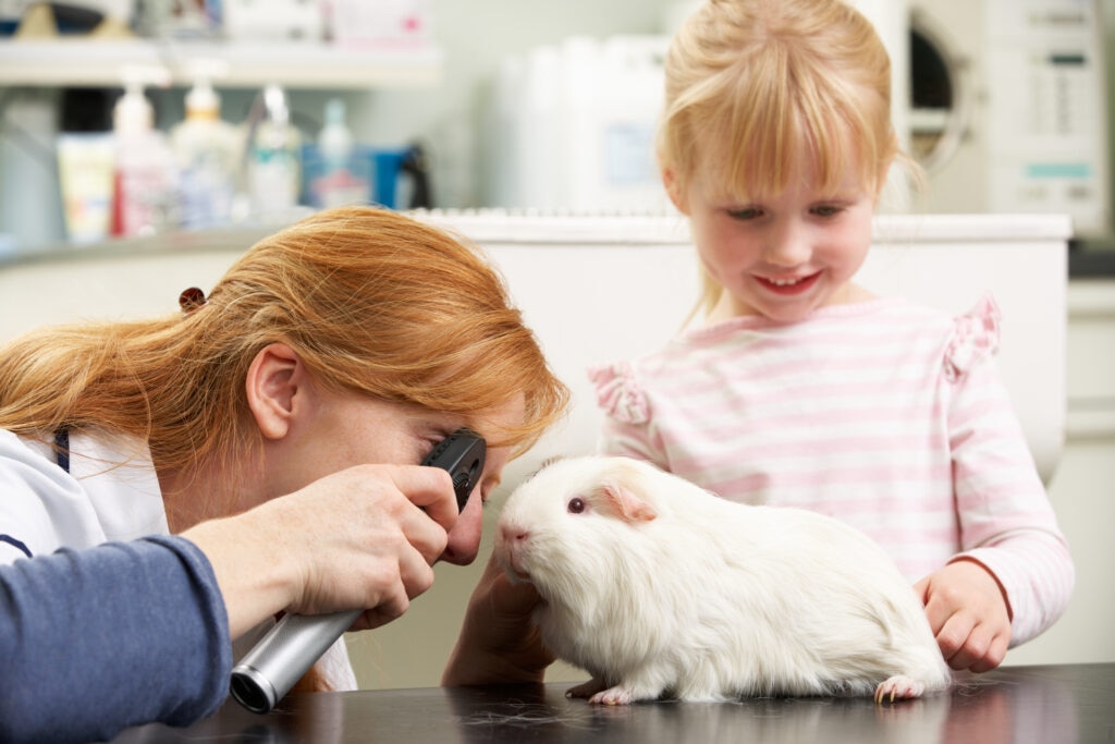
Eye exam: Uses an ophthalmic scope that has a light and magnifying lens to look closely at the eyelids, cornea, conjunctiva, retina, and surrounding tissues
-
Fluorescein stain: A special green dye is placed on the eye, and if there is an ulcer or a scratch, the damaged area will absorb the dye, which helps make the diagnosis.
-
Tear testing: Used to assess if tear production is normal either via Schirmer tear test or phenol red test. Both are placed on the lower eyelid for 15 seconds and the strip or string turns red or blue from the tears produced. If the tears make it to20mm on the strip or string in 15 seconds, that is typically a normal number of tears.
-
Referral exam: Severe ulcers may need to be checked by a veterinary ophthalmologist.
Before the appointment or during, let your veterinarian know when the symptoms started, how the eye has looked since it started, if there have been any appetite or stool changes, what kind of bedding and hay you use, any medications or supplements you have tried so far to help, and if there have been any fights with cage mates.
Taking photos of the eye each day and showing your vet can be extremely helpful.
Treatment of Corneal Ulcers in Guinea Pigs
Treatment depends on the ulcer’s cause and depth, and if there is an infection. Corneal ulcers are often very treatable, but they need veterinary care and close monitoring. Simple ulcers can heal in seven to 14 days; more complex, deep, or infected ulcers can take much longer.
Treatment can include:
-
Removing any foreign material stuck in the eye, like hay or hair. Your vet may flush the eye to gently remove the object.
-
Topical antibiotic eye medication to treat infection or help prevent the ulcer from becoming infected
-
Lubricating eye ointment or drops to help protect the eye surface
-
Pain control
-
Treatment for any eyelid or hair problems. If the eyelid anatomy is abnormal and rubbing on the cornea, surgical correction of the eyelid or removal of hair can help.
Deep, infected, or melting ulcers are often treated by an ophthalmologist and can need surgery and possible around-the-clock monitoring in the hospital for eye drop treatment depending on severity.
Recovery and Management of Corneal Ulcers in Guinea Pigs
A mild, superficial ulcer may heal in about one to two weeks, while deeper, infected, or nonhealing ulcers may take several weeks with advanced care.
During recovery, take these steps:
-
Make sure to give all medications as prescribed.
-
Avoid touching the medication tip to the eye, eyelids, fur, or skin.
-
Keep the habitat clean and dry.
-
Switch to low-dust, soft bedding.
- Wash all fleeces and liners.
-
Monitor appetite, stool output, energy levels, and activity.
Contact your veterinarian right away if the eye looks cloudier, swollen, painful, red, or enlarged. Also call them if your guinea pig stops eating, is less active, or has decreased stool production.
Prevention of Corneal Ulcers in Guinea Pigs
Not all corneal ulcers can be prevented; guinea pigs are at higher risk due to their low tear production in general.
To minimize their risk, use soft, low-dust bedding and avoid cedar shavings or dusty bedding. Keep the enclosure clean and do deep cleans weekly.
Remove any broken toys or sharp areas of the cage. Keep guinea pigs in separate cages if they start fighting. It’s also important to schedule annual veterinary exams, especially if your guinea pig has any eye discharge or has had corneal ulcers in the past.
Corneal Ulcers in Guinea Pigs FAQs
How do you treat corneal ulceration in guinea pigs?
Treating a corneal ulcer in guinea pigs starts with a thorough eye exam by a veterinarian.
Treatment may include prescription antibiotic eye medication, eye lubrication, pain control, and changes to the enclosure to reduce any irritation.
Severe cases often involve intensive treatment by a veterinary ophthalmologist.


