Histiocytoma in Dogs: Symptoms and Treatment


Ridofranz/iStock/Getty Images Plus via Getty Images
Histiocytomas in dogs are common, benign skin masses that typically appear as single, raised red bumps. They often regress on their own within a few months, but a veterinarian should examine any new lump to rule out more serious skin tumors.
Key Takeaways
- Histiocytomas are common, benign skin masses that often appear as single, raised red bumps.
- They usually regress and disappear on their own within a few months.
- Veterinary diagnosis is important because more serious skin tumors can look similar to histiocytomas.
- Treatment may be needed if a histiocytoma becomes irritated or infected, doesn't regress, or causes discomfort.
What Is a Histiocytoma in Dogs?
Cutaneous histiocytomas (CCH) in dogs are fairly common benign (non-cancerous) skin masses that are not usually a cause for concern.
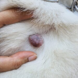
Sometimes referred to as button tumors in dogs, histiocytomas appear as single red bumps that are generally less than an inch wide. They most commonly grow on a dog's head, face, ears, or legs.
These pink growths usually have smooth surfaces but can become open wounds if a dog licks the area repeatedly.
Histiocytomas can grow quickly early on, but they typically start to regress and disappear within a few months. Proper diagnosis by a veterinarian is important to differentiate histiocytomas from other more serious skin tumors that can look similar.
Histiocytomas are common in dogs of all ages but particularly in dogs 3 years of age or younger. Boxers, Boston Terriers, and Labrador Retrievers are a few of the breeds most commonly affected.
If you notice any lumps or bumps on your dog's skin, schedule an appointment with your veterinarian for a proper diagnosis and to discuss suitable treatment options.
What Are the Symptoms of Histiocytomas in Dogs?
Histiocytomas in dogs generally do not cause alarming symptoms and typically appear as a hairless, raised, red skin bump. Pet parents often first notice the bump while petting or grooming their dog.
Signs of histiocytomas in dogs may include:
- Hairless, raised, red skin bump
- Bleeding
- Itching
- Open sore with pus, if infected
- Swelling around the lump, if infected
What Causes Histiocytomas in Dogs?
The exact underlying cause of histiocytomas in dogs isn't fully understood.
Because histiocytomas are made up of a special type of immune cell, it's likely that dysregulation within the immune system plays an important role.
There may also be a genetic component because several breeds are at an increased risk of developing histiocytomas.
How Are Histiocytomas in Dogs Diagnosed?
Veterinarians diagnose histiocytomas in dogs with a thorough physical exam and may use a fine-needle aspirate (FNA) or biopsy to confirm the diagnosis.
Your veterinarian will first perform a thorough physical exam, paying close attention to your dog's skin. If a bump is found that the veterinarian suspects may be a histiocytoma, testing can be done to confirm the diagnosis.
Tests may include:
- Fine-needle aspirate (FNA): A veterinarian gently inserts a needle into the skin mass to collect a sample of cells. The sample is placed onto a glass slide, stained, and viewed under a microscope to identify the type of cells present.
- Biopsy: A small portion of the mass—or the entire mass—is removed and sent to a laboratory for special testing. This is generally done after an FNA for a more definitive diagnosis and requires local anesthesia or sedation.
How Are Histiocytomas in Dogs Treated?
Most histiocytomas in dogs don't require treatment. They usually regress on their own within three months as the immune system controls their growth.
Treatment for Irritated or Infected Histiocytomas
If a histiocytoma is in an area with frequent contact or your dog repeatedly licks or scratches it, the mass may bleed and become infected. This is quite common for histiocytomas on a dog's paw.
In these cases, topical or oral antibiotics such as Animax® ointment or cephalexin may be indicated. These typically cost $10–$15.
Surgical Removal
If the histiocytoma doesn't regress or causes discomfort, surgical removal may be recommended by a veterinarian for a permanent solution.
If surgery is needed, costs generally range from $300–$500, but this is highly variable based on where you live. If a biopsy is sent to the laboratory, additional costs may be around $200–$400.
Recovery and Management of Histiocytomas in Dogs
Histiocytomas generally do not cause discomfort. However, histiocytomas on a dog's legs may rub against the ground and become irritated.
An orthopedic dog bed can help cushion these areas and prevent irritation. Some dogs may also need a recovery cone to prevent them from licking or itching the area.
If you notice bleeding or oozing from the histiocytoma, bring your dog to the veterinarian to rule out an infection.
How To Prevent Histiocytomas in Dogs
Because the cause of histiocytomas isn't fully understood, it's difficult to determine the best ways to prevent them.
Histiocytomas are generally not harmful to a dog's health, so pet parents can focus on preventing irritation and infection while waiting for the growths to regress. This includes closely monitoring the histiocytoma and promptly bringing any concerns to your veterinarian.
Histiocytoma in Dogs FAQs
Is there a home treatment for histiocytomas in dogs?
No. There are no home remedies to treat histiocytomas in dogs. They almost always spontaneously regress on their own.
While the histiocytoma regresses, closely monitor your dog for signs of irritation or infection.
How quickly do histiocytomas grow?
Histiocytomas in dogs can grow quickly within the first month and may reach around 1 inch in width during this stage.
How can you tell the difference between a mast cell tumor and a histiocytoma in dogs?
It can be difficult to tell the difference between a mast cell tumor and a histiocytoma based on appearance alone because the two skin masses may look similar.
A veterinarian must perform a fine-needle aspirate (FNA) or biopsy to diagnose the skin mass.


