Ultrasounds for Pets: What To Expect


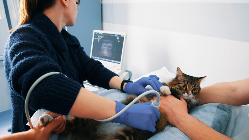
alexey_ds/E+ via Getty Images
An ultrasound is a noninvasive type of diagnostic imaging. Ultrasound is commonly used in dogs and cats but is also an important diagnostic tool for many exotic pets, including rabbits, ferrets, guinea pigs, birds, and reptiles.
Key Takeaways
- Pet ultrasounds use high-frequency sound waves to create real-time images of organs and soft tissues, helping veterinarians diagnose many conditions without using radiation.
- Veterinarians may recommend an ultrasound to evaluate symptoms or conditions such as vomiting, abdominal pain, weight loss, pregnancy, heart disease, or suspected internal abnormalities in dogs, cats, and many exotic pets.
- Most pet ultrasounds are painless, take 20–60 minutes, and do not require sedation unless a pet is anxious, painful, or needs a biopsy or fluid sample collected.
What Is an Ultrasound for Pets?
With an ultrasound, veterinary staff get a real-time view of a pet’s organs and soft tissues using high-frequency sound waves.
These images or videos are used to diagnose issues affecting internal organs or tissues.
There are several differences between X-rays and ultrasound.
- X-rays expose the pet to radiation while ultrasound does not.
- X-rays take a still image while ultrasound can be used to look at organs or tissue in motion, such as the heart while beating.
- X-rays are better at showing bones while ultrasound is better for soft tissue and organ detail.
- Some ultrasound machines can display blood flow.
Why Would a Pet Need an Ultrasound?
Common Reasons for Dogs and Cats
There is a plethora of reasons that a dog or cat might get an ultrasound, including:
- Vomiting, diarrhea, or weight loss
- Abdominal pain
- Abnormalities of the liver, kidneys, bladder, reproductive tract, or spleen
- Pregnancy evaluation
- Heart disease (with an echocardiogram, which is an ultrasound of the heart)
- Blood in urine
- Checking for internal bleeding
- Diagnosis or staging of cancer
- Lung exam in patients with respiratory signs
- Guidance for the veterinarian to collect biopsy or fluid samples
Common Reasons for Exotic Pets
Examples of reasons an exotic animal might get an ultrasound include:
- Egg binding or reproductive tract concerns
- Pregnancy evaluation in mammals
- Liver or kidney disease
- Gastrointestinal blockages or other issues
- Cancer, masses, or unexplained swelling
- Checking for fluid under the shell of turtles or tortoises
- Checking the heart
- Weight loss or changes in droppings
What Happens During a Pet Ultrasound?
Most ultrasounds are painless and take 20–60 minutes to complete, depending on the animal’s size and what part of the pet is being examined.
Step by Step
- The veterinarian will do a physical exam and review your pet’s medical history.
- If necessary and safe, the pet is sedated. Most ultrasounds can be done without sedation.
- Fur may be clipped over the imaging site if necessary and applicable. In pets with feathers, the feathers are usually parted or dampened to allow the probe to touch skin.
- The pet is positioned comfortably depending on what part of their anatomy is getting scanned.
- Ultrasound gel is put on the skin or the ultrasound probe.
- The veterinarian or veterinary radiologist does the ultrasound scan.
- The images will be interpreted (read and analyzed) and next steps discussed.
If there is a mass or abnormal fluid buildup in the abdomen, the veterinarian may recommend collecting a sample. Sometimes fluid samples can be taken without sedation, but sedation is recommended for sampling of organs to make sure the pet does not move.
How To Prepare Your Pet for an Ultrasound
Before the Appointment
- Check with your veterinarian to see if fasting (withholding water and/or food) is recommended and then follow their fasting instructions.
- Give medications as directed by the veterinarian. If sedation or anesthesia will be used, ask which of your pet’s usual medications are safe to give.
- If asked, bring previous imaging or medical records with you.
- If your pet is stubborn, aggressive, or anxious, the veterinarian may recommend antianxiety medications be given before the appointment. Give these as directed.
Species-Specific Considerations
- Dogs: The veterinarian may recommend fasting your pet and taking them outside to poop or pee before the appointment if the abdomen is being scanned.
- If the bladder is being scanned, you may be told to prevent urination.
- For aggressive or stressed pets, anti-anxiety medications may be recommended.
- Cats: You want to make sure that your cat arrives in a good mood for their ultrasound scan. Consider the use of feline calming pheromones, such as Feliway spray, within the cat’s carrier. Work to get your cat used to the carrier before the appointment.
- Give antianxiety medications as recommended by your vet. You may consider clipping claws to prevent scratches as well. Check with your veterinarian for fasting recommendations.
- Exotic pets: Fasting recommendations vary widely by species. Only fast your exotic pet if instructed to do so by your veterinarian.
- Consider how to keep the right temperature and reduce stress during transport. This may involve driving a temperature-controlled vehicle, using heat packs or insulated carriers, or covering cages or carriers with breathable blankets.
- Make sure to check with your veterinarian about whether you need to do anything different with the pet’s care leading up to the appointment.
Will My Pet Need Sedation?
Most pets do not require sedation for routine ultrasonographic examination. Sedation may be necessary if:
- The pet is aggressive
- Your pet has too much pain or anxiety to remain still
- A guided biopsy of a tissue sample will be taken
If sedation is needed, your veterinarian will decide the safest option for your pet based on their species and individual health.
What Can an Ultrasound Diagnose?
Ultrasound is often used with other diagnostics, such as blood work or biopsy, to figure out a pet’s disease or condition. Ultrasound can play a key role in diagnosing many health conditions, such as:
- Liver disease
- Kidney disease
- Bladder stones or abnormalities
- Pancreatitis
- Masses and tumors
- Fluid gathering within the abdomen or chest
- Gastrointestinal disease or blockage
- Heart disease
- Egg binding in reptiles and birds
- Pregnancy in mammals
Are Ultrasounds Safe for Pets?
Ultrasounds are considered very safe for pets.
This diagnostic does not use radiation. Risks associated with ultrasound do not come from the technology itself. Rather, risks are from anesthesia, sedation, or the collection of tissue or fluid samples.
Ultrasounds for Pets FAQs
How long does a pet ultrasound take?
Most ultrasounds take 20–60 minutes depending on the pet’s size and what part of their body is being scanned.
Does an ultrasound hurt my pet?
Ultrasound is usually not painful. Pets who have pain in their abdomens or other areas may be uncomfortable with the probe pressed against the painful spot.
Will my pet need anesthesia during an ultrasound?
Most pets do not need anesthesia during an ultrasound. Sedation may be needed for animals who are aggressive, anxious, or in pain. Sedation may also be necessary if a tissue sample needs to be taken.
Can exotic pets have ultrasounds?
Yes, exotic pets can have ultrasounds. Many vets, however, are not trained in exotic pet ultrasound, so you may need to call around to find someone willing and able to do this diagnostic for your pet.
How soon will I get the results from an ultrasound for my pet?
You may get results the same day. If the veterinarian needs to have them interpreted by a radiologist, results may take a day or two.
What is the difference between an ultrasound and an X-ray for pets?
Ultrasound uses high-frequency sound waves rather than radiation to make images of organs and soft tissues. Images are captured in real time and can be viewed like a video. X-rays use radiation to make snapshots, so the pet is exposed to radiation, and you only get a still image. X-rays are better for imaging bones.


