Microscopic vs Macroscopic Disease — What is the Difference?


Now that the Cardiff has recovered from two surgeries to remove an intestinal tumor and multiple skin masses, it's time move onto the topic of treating cancer that could still be lurking in his body.
Surgery to cut out the area of T-Cell Lymphoma on his small intestine was successful in alleviating his clinical signs of vomiting, diarrhea, decreased appetite, and lethargy. Removing the tumor and not being able detect any cancer cells in other body tissues essentially put him into remission. Unfortunately, there still is the chance that cancer cells are present in his body that will form new tumors.
Comparing cancer that can be seen or palpated (touched) to that which may not have yet developed to a detectable size comes down to differentiating between microscopic and macroscopic disease.
What is Microscopic Disease?
Microscopic disease is the level of cellular change that can't be seen with the naked eye. That is, the disease can be visualized on a slide under the microscope but can't otherwise be readily detected.
With cancer, microscopic disease occurs all the time, as cells having abnormal DNA divide rapidly without the proper mechanism to turn off their division.
It's takes days to months for enough cancer cells to divide to create a tumor that can then be discovered through physical examination, diagnostic testing, or through the development of clinical signs of illness. As a result, even though your veterinarian may not be able to find cancer in your pet at a particular time, the potential exists that cancer cells that ultimately will form tumors are existing in the body.
An image of Cardiff’s microscopic disease as seen on the biopsy of his intestinal tumor is provided courtesy of Idexx Laboratories and is included at the end of this column.
What is Macroscopic Disease?
Macroscopic disease is that which your veterinarian can discover on a physical examination or via diagnostic techniques like radiographs (x-rays), ultrasound, CT, or MRI.
As Cardiff’s cancer returned 12 months after completing his first course of chemotherapy in July 2014, either his chemotherapy didn't kill all of the cancer cells (microscopic disease) or he is just prone to forming cells containing abnormal DNA that then divided without stopping and ultimately formed a tumor. The second option is most likely, as he had such a long disease-free interval, especially considering the poor prognosis that goes with T-Cell Lymphoma.
As development of macroscopic disease can happen so fast in some patients, it’s crucial to follow your veterinarian’s guidelines for rechecks, which normally include physical examination, and diagnostics like blood and urine testing, radiographs, ultrasound, and others tests.
Cardiff had been getting blood testing every one to four weeks and chest radiographs and abdominal ultrasound every three to four months. Yet, the development of a new intestinal mass occurred and wasn’t seen in the short period of one month between his prior ultrasound in June 2015 and the most recent ultrasound in July 2015.
When Cardiff was again diagnosed with an intestinal mass, we were able to visualize his macroscopic disease via ultrasound. Cardiff’s surgeon, Dr. Justin Greco of ACCESS LA, was able to both see and feel the mass during the exploratory abdominal surgery.
When it comes to treating macroscopic disease, surgically removing it is most ideal for the patient, provided the patient is healthy enough to endure the anesthesia and surgery.
“A chance to cut is a chance to cure” rings true and can alleviate clinical signs of disease or potentially change the course of treatment in a favorable way, such as putting the patient into remission and reducing the need for chemotherapy or radiation to treat residual disease.
Removing only a section of a tumor from the body via surgery is still helpful, but leaving behind cancer cells increases the need for chemotherapy and radiation to manage the residual disease.
An image of Cardiff’s macroscopic disease, as seen after his intestinal tumor was removed, is also included at the end of this article.
What Does This Mean for Cardiff?
Even though Cardiff had surgery which completely removed his intestinal tumor with wide margins, there's still a chance that he has microscopic disease lurking elsewhere in his body.
I feel confident that surgery resolved Cardiff’s macroscopic disease, but Cardiff’s veterinary oncologist, Dr. Avenelle Turner, and I still feel that it's best to put him through a course of chemotherapy to kill any microscopic a disease. Doing so will decrease the likelihood that he’ll develop further microscopic and macroscopic disease in the future.
Check back next time as I discuss Cardiff’s chemotherapy plan and give an update on his status.
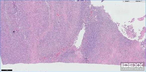
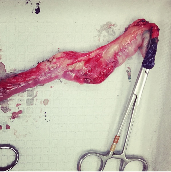

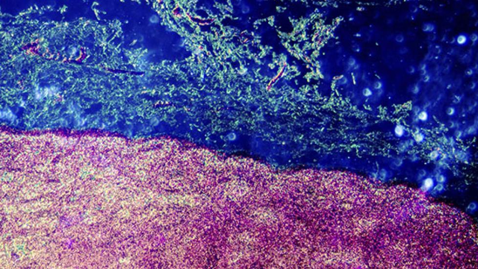
Image: Lymph gland tissue / Thinkstock
Related
When Cancer That Was Successfully Treated Reoccurs in a Dog
What Are the Signs of Cancer Reoccurence in a Dog, and How is it Confirmed?
Surgical Treatment of Canine T-Cell Lymphoma in a Dog
What We Do When There Are Tumors On the Inside and On the Outside
What Makes One Skin Mass Cancerous and Another Non-Cancerous?


