Acute Liver Failure in Cats: Signs and Treatment


FreshSplash/E+ via Getty Images
Liver failure in cats is a significant and life-threatening medical condition, especially if left untreated.
This article explores its symptoms, causes, and treatment options so you are better prepared to care for your cat.
Key Takeaways
- Acute liver failure in cats is a life-threatening medical condition and veterinary emergency.
- Symptoms of acute liver failure in cats may include decreased appetite, vomiting, and diarrhea.
- In cases of end-stage liver failure, after managing complications and keeping quality of life for as long as possible, unfortunately, humane euthanasia may be necessary.
What Is Acute Liver Failure in Cats?
Acute liver failure in cats occurs when there suddenly isn’t enough liver tissue for the organ to carry out its normal and essential functions.
The liver absorbs and makes multiple proteins and clotting factors, neutralizes and gets rid of wastes and toxins, controls glucose (sugar) levels, and aids in daily digestion and metabolism processes.
Because of its functions, the liver is at high risk for damage from a variety of sources, but fortunately, it has the unique ability to regrow after it’s damaged. Studies have shown that liver failure happens when more than 70% of the liver is damaged.
Acute is a vague term, but usually, symptoms are shown within a few days of initial damage.
As a result, acute liver failure is often harder to reverse and results in a higher death rate. Some causes of liver failure can be cured if found and treated early enough.
Acute liver failure in cats is considered a medical emergency and is a painful and exhausting condition that can result in death.
Cats with end-stage liver failure (cirrhosis) often die within days of diagnosis. For cats that recover, long-term liver damage can occur, leading to scarring of the liver and ultimately to cirrhosis.
Symptoms of Acute Liver Failure in Cats
Symptoms of acute liver disease in cats are often vague and can take time to be noticeable. Many pets with this condition have:
-
Abdominal pain
-
Weakness
As the disease progresses and greater liver damage happens, symptoms will become more severe, such as:
-
Bleeding from the nose, mouth, rectum, and bladder
-
Collapse
-
Icterus (yellowing of the skin, gums, and eyes)
-
Melena (black tarry stools)
-
Neurologic and behavioral changes such as: head pressing, seizures, circling, lack of coordination, hyperactivity, excessive salivation, blindness, or coma
-
Petechiae and ecchymoses (bruising noted on the skin, gums, and eyes)
Causes of Acute Liver Failure in Cats
In cats with acute liver failure, sometimes the underlying cause may be unknown (termed idiopathic), but there are several known causes of liver failure, such as:
-
Abscess, which results from umbilical cord infections in kittens but can also occur in adult cats from bacteria such as Yersinia pestis and Actinomyces spp.
-
Cancer like lymphoma
-
Cholangiohepatitis or triaditis, inflammatory disease often affecting the gallbladder, pancreas, and liver at the same time
-
Heart failure and clots
-
Heavy metals like copper, although rare in cats
-
Hepatic lipidosis, which is the most common and potentially reversible cause of acute liver failure
-
Infectious organisms: Clostridium (bacterial), histoplasmosis (fungal), liver flukes (parasitic), toxoplasmosis (protozoal), feline infectious peritonitis and FeLV/FIV (viral)
-
Inherited conditions such as amyloidosis—seen in breeds like Abyssinians, Siamese, and other Oriental cats—and biliary cystic disease, more common in Persians
-
Medications such as diazepam and Tylenol (acetaminophen)
-
Portosystemic shunts, usually a congenital issue (present at birth)
-
Toxins such as blue-green algae, mycotoxins (aflatoxin, usually through eating moldy or spoiled food), rodenticides, pine oil
How Veterinarians Diagnose Acute Liver Failure in Cats
Cats can get acute liver failure from many causes. Because the symptoms are often vague, diagnosing acute liver failure can be difficult.
Veterinarians rely on tools such as blood work and urinalysis in addition to a physical exam to diagnose acute liver failure in cats.
After these basic tests, your veterinarian may need to perform additional tests to check liver function and severity of liver damage.
For a more accurate diagnosis, a liver sample is needed, which can be taken either through surgery or biopsy, where a needle is put directly into the liver.
Treatment of Acute Liver Failure in Cats
Because cats with acute liver failure are very sick, hospitalization of several days is often needed.
The goal of treatment for acute liver failure in cats is to slow the disease progression and allow time for the liver to regrow.
Therapy for the underlying cause, if that cause is known, is ultimately needed along with early and aggressive symptomatic treatment, including the following:
-
Antacids, antinausea medications (Cerenia, famotidine, ranitidine), and gastroprotectants like sucralfate
- IV fluid therapy (and/or blood or plasma transfusions) with electrolyte and glucose supplementation
-
N-acetylcysteine
-
SAMe and milk thistle derivatives (silymarin) along with ursodiol
-
Pain control
-
Flumazenil for suspected cases of diazepam intoxication.
-
Vitamin K1 to aid with clotting
For known or suspected cases of eating or drinking a toxin, talking with Pet Poison Hotline (855-764-7661) is advised. Consultation fees may apply.
Dietary therapy can also help with liver function and slow further damage to the liver.
Prescription diets such as Hill's I/D, Royal Canin hepatic, or Purina HP Hepatic have more digestible protein, more antioxidants that help protect cells from damage, and, to help with fluid levels, lower amounts of sodium.
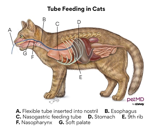
For some cats, especially those with hepatic lipidosis, feeding tubes are needed to deliver nutrients. Be sure to discuss the kind of diet, if applicable, with your veterinarian.
As some cats with acute liver failure may have neurologic signs, daily nursing care—such as frequent turning them over onto their other side, soft bedding to prevent decubital ulcers, and keeping them dry and clean—is important. Enemas, lactulose, and antibiotics like neomycin or metronidazole may be needed in these cats as well.
In cases of end-stage liver failure, after managing complications and keeping quality of life for as long as possible, unfortunately, humane euthanasia may be necessary.
Recovery and Management of Acute Liver Failure in Cats
If the underlying cause is treated early and aggressively enough, some cats can recover and have a good quality of life.
This is largely due to the liver's ability to regenerate.
Prevention of Acute Liver Failure in Cats
Finding the issue early and having routine veterinary check-ups can go a long way to ensuring a positive outcome.
Not all causes of liver failure can be prevented, but there are some ways to decrease your cat’s risks.
You can:
-
Keep garbage and medications away from your cat’s reach.
-
Block access to medications and toxins, like rodenticide.
-
Keep your cat vaccinated and routinely dewormed.
-
Take steps to avoid sources of possible bacterial contamination by keeping your cat indoors.
-
Have your cat examined right away if she stops eating.
-
Talk to your veterinarian about the risks and benefits of certain medications for your cat.
Acute Liver Failure in Cats FAQs
Can the liver recover from acute liver failure?
Fortunately, the liver has a large reserve capacity and there are many conditions it can recover from.
How do you treat acute liver failure in cats?
Treatment of acute liver failure in cats is largely symptomatic and supportive care (e.g., fluid therapy, antibiotics, antioxidants, gastroprotectants) to relieve pain and discomfort while giving the liver time to recover.
Ultimately, treatment is aimed at the underlying cause, if found.
What does end-stage liver failure look like in cats?
Cats with end-stage liver failure are truly sick.
They will have yellowing of the skin and gums, and they often have pinpoint hemorrhages (petechiae) throughout their body and swollen limbs and belly; will act depressed or lethargic; or are in a coma.
How long will a cat live with liver failure?
Cats with liver failure have different outlooks depending on the underlying cause.
With prompt treatment, acute liver failure can be managed, giving the liver time to recover and for those cats to go on with a normal life.
Other diseases, like cancer, or complications that can arise from acute liver failure can result in a poor quality of life and short lifespan.
References
Cullen JM. Summary of the World Small Animal Veterinary Association standardization committee guide to classification of liver disease in dogs and cats. Veterinary Clinics of North America: Small Animal Practice. 2009;39:395-418. https://doi.org/10.1016/j.cvsm.2009.02.003.
Hughes D, King LG. The diagnosis and management of acute liver failure in dogs and cats. Veterinary Clinics of North America: Small Animal Practice. 1995;25(2):437-460. https://doi.org/10.1016/s0195-5616(95)50036-1.