Keratomas in Horses


DebraCarrPhotography/iStock / Getty Images Plus via Getty Images
What Are Keratomas in Horses?
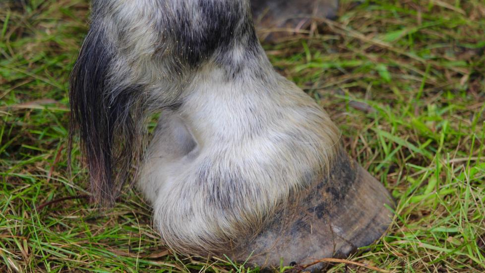
Keratomas are benign (non-cancerous) growths that occur within the hoof of the horse. While relatively uncommon, keratomas can be uncomfortable for the horse and time-consuming to manage if not addressed promptly.
The hoof is a fixed capsule, meaning it can’t expand as a keratoma grows, so the growth will push on important surfaces within the foot, such as:
-
Bones
-
Tendons
-
Joints
-
Soft tissue structures
Keratomas in horses may cause significant lameness as the mass grows.
There are no breed, age, or gender predispositions for a horse to develop keratomas, so any horse may be susceptible to developing them.
Symptoms of Keratomas in Horses
Some common clinical signs of keratomas in horses include:
-
Frequent hoof abscesses
-
Bulging of the coronary band or hoof wall
-
Toe-pointing behavior when standing to take pressure off the foot
-
Thickening or displacement of the white line
Causes of Keratomas in Horses
The hoof wall is partly made up of a protein called keratin, which is produced by keratinocytes. Keratomas occur when there is an abnormal growth of keratoma inside the foot.
Horses with previous trauma to the coronary band or hoof may be more predisposed to keratomas, although this is not always a direct result. However, the reason why extra keratin is made in some horses and leads to keratomas is still not exactly known.
As keratomas in horses grow larger, they often cause a separation in the hoof wall; this allows for bacteria to enter the foot and can cause foot abscesses.
As the keratoma increases in size, it encroaches on the coffin bone in the foot and creates a defect, which causes a bulge to appear.
How Veterinarians Diagnose Keratomas in Horses
Veterinarian will typically begin diagnosis with a basic lameness exam. This includes:
-
Physical examination
-
Observing the horse move at the walk and trot
-
Using hoof testers to check for any sensitivity in the foot
-
Using nerve blocks to help isolate the source of lameness
If lameness is found in the foot, your veterinarian will likely take X-rays. This will allow your veterinarian to evaluate the anatomy of the internal structures of the hoof such as:
-
Phalanx bones
-
Joint space widths
-
Angles of the hoof wall and coffin bone
-
Sole depth
Keratomas in horses appear on X-rays as a dark “divot” in the third phalanx (coffin) bone.
Your veterinarian may recommend further diagnostics like a CT scan if the X-ray imaging is not conclusive.
Treatment of Keratomas in Horses
The curative treatment for keratomas in horses is surgical removal via hoof wall resection, which means cutting open the hoof wall and making a hole wide enough to scrape and scoop out the mass.
This procedure is either done with the horse standing and sedated with nerve blocks to “deaden” the foot for surgery, or with the horse under general anesthesia. The surgical method is dependent on the horse’s attitude, level of keratoma involvement, and veterinarian comfort level.
Recovery and Management of Keratomas in Horses
Following surgery, the affected hoof is bandaged for several days with an antibiotic-soaked gauze placed in the surgical site.
Horses are usually prescribed a systemic antibiotics like SMZ-TMP and anti-inflammatories such as phenylbutazone through initial recovery.
After the hoof bandage is removed, your veterinarian will likely work with your farrier to have a hospital plate or egg-bar shoe with clips applied to minimize movement of the hoof wall for several weeks. During this time, the hoof will develop sufficient granulation tissue or horn development of the wall to be able to withstand full pressure while weight-bearing.
The full defect of the hoof wall surgical site may take several months to grow out completely. With appropriate treatment, long-term prognosis for horses with keratomas is excellent.
Prevention of Keratomas in Horses
Because all horses are susceptible to potentially developing keratomas, there is no “prevention.” Most horses only develop one keratoma, although there is a possibility for development in multiple feet or to have more than one mass in the same foot. The best thing you can do for your horse is have them immediately examined by your veterinarian if you notice any lameness or bulging of the hoof wall or coronary band.
Keratomas in Horses FAQs
Can a horse live with a keratoma?
While a keratoma is a benign growth, it can lead to severe discomfort, chronic lameness, and repeat abscesses. This may lead to other secondary conditions like laminitis, weight loss, and depression due to the chronic pain.
What is the prognosis for a horse with a keratoma?
After an accurate diagnosis has been made, the prognosis for keratomas is good to excellent if medical intervention is addressed early.
How do you know if your horse has a keratoma?
A keratoma is diagnosed by a lameness localized to the foot, followed by diagnostic imaging such as X-rays.