What Is a Horse Hoof Made Of?


Horse Hoof Anatomy
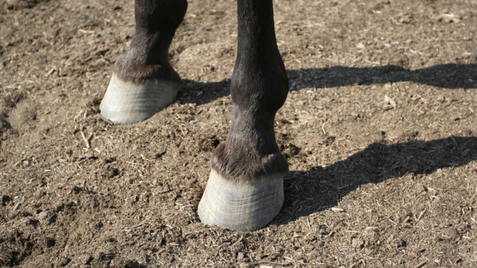
A horse’s hoof can be thought of in three parts: the outside or wall, the inside functional structures, and the weight-bearing surface.
The Hoof Wall
The outside of the hoof, also known as the wall, is the part of the hoof you see when the horse is standing which is responsible for bearing weight. From front to back the hoof is split into sections called the toe, the quarter, and the heel. The hoof wall is a hard surface, made of a durable protein called keratin that protects the foot and is comprised of three layers.
-
The outer layer is the hardest part of the hoof and is responsible for protecting the more vulnerable inner structures. It does not contain nerves or vasculature (blood vessels).
-
The middle layer is the thickest part of the hoof and is responsible for rigidity and structure.
-
The inner layer is comprised of laminae (finger-like projections) that interlock with the laminae covering the coffin bone (P3 or third phalanx) inside the hoof to keep it in place.
Inflammation of these laminae (from carbohydrate overload, stress, systemic illness, or other infection) is called laminitis and can lead to rotation or sinkage of P3 and may cause severe, sometimes irreversible, lameness.
The coronary band is responsible for the growth and nutrition of the hoof. It is the ring at the very top of the hoof where it meets the skin/coat of the pastern. The coronary band writes the horse’s history subtly, showing things like major dietary changes, stress, and laminitic episodes by changes in coloring, hardness, flaking, and lines as it forms and grows out.
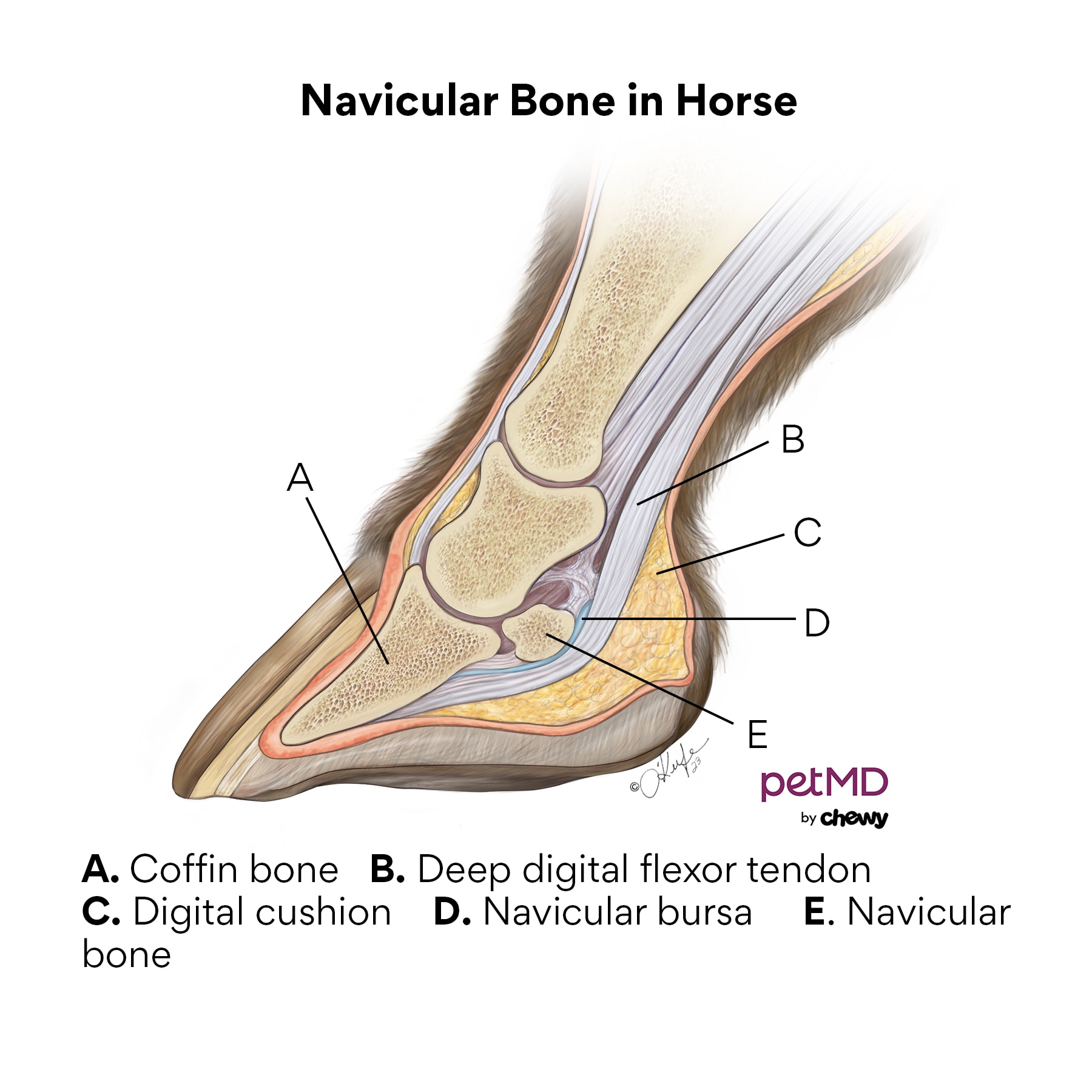
Internal Structure
The internal structures of the foot include:
-
Second and third phalanx
-
Lateral cartilages (“wings”) of P3t
-
Navicular bone and bursa
-
Deep digital flexor tendon (DDFT) and other ligaments
-
Joint spaces
-
Countless blood vessels, nerves, and lymphatic structures
Weight-Bearing Surface
The weight bearing surface, or bottom, of the hoof is comprised of several parts:
-
The hoof wall is the tough outer ring. Along with the bar and frog, this is the actual weight-bearing surface of the foot.
-
The bars turn in from the heels, surround the frog, and run towards the point of the toe.
-
The frog is a softer, almost rubbery-feeling triangular structure in the center of the foot responsible for blood flow and circulation each time the horse steps.
-
White line is an accurate term for the typically thin line that depicts the transition from hoof wall to the sole.
-
The sole is the wide, slightly soft, and concave structure making up most of the bottom of the foot.
-
The digital cushion helps form the heels and is responsible for shock absorption and blood flow through the foot along with the frog.
Is a Horse Hoof Like a Fingernail?
Both the fingernail and horse hoof are comprised of a hard protein called keratin. Like fingernails, the hoof wall grows continually, at a rate of ¼–½ inch per month, so a horse can grow a whole new hoof in approximately a year.
This is similar to how human nails will have white spots or lines from bruising or other traumatic events. Both will break off and need trimming as they get long. In horses, this will prevent excess tension and stress along the flexor tendons and internal laminae and keep the foot balanced. While we use fingernail clippers, farrier tools are a bit heftier to cut through and rasp or file the thicker surfaces.
Are Hooves Painful for Horses?
While the hoof wall itself does not feel pain due to its lack of blood supply and nerves, its connections to internal structures and the coronary band (where the wall originates) are sensitive.
Pain experienced from the more sensitive structures could be due to:
-
Inflammation (from instances of laminitis, stone bruises, etc,
-
Trauma (major lacerations, stepping on a nail)
-
Degenerative joint disease/arthritis
-
Navicular disease
-
Soft tissue injuries to flexor tendons
-
Other conditions
When picking debris out of the hoof, it’s important to be careful around the heel bulbs, frog, and sulci, the softer parts of the hoof, as they can be more sensitive than others. These areas are softer, therefore more susceptible to pressure, which can damage underlying vessels and tissue like the laminae.
When applying shoes correctly, a nail is hammered through the hoof wall from the bottom surface of the foot, out to the hoof wall at an angle that avoids any of the laminae or sensitive tissue. If done incorrectly or if the hoof’s internal anatomy varies, this can lead to pain, inflammation, or infection of the very sensitive laminae.
What Is the Bottom of a Horse’s Foot Made Of?
As mentioned above, the bottom of the foot is comprised of the hoof wall, white line, sole, bars, and digital cushion. The frog and the digital cushion (part of what forms the heels, and the most elastic part of the foot) are more “springy” materials responsible for shock absorption each time the foot hits the ground as well as blood circulation.
Each time the horse takes a step, these structures flatten out, slightly widening the bars and compressing the vasculature (veins and arteries) within the bottom of the foot. As the horse picks that foot up, the vessels bounce back elastically like a coil that had been stretched to push blood up and out of the hoof working against gravity. This is what allows for adequate blood flow to the lowest part of the limb, keeping these structures vital and promoting healthy hoof growth.
What Is the Most Sensitive Part of the Horse’s Hoof?
Technically the most sensitive part of the hoof is its attachment to internal structures. The frog and digital cushion/heel bulbs are also sensitive, as they are softer and play a large role in circulation.
Problems with Horse Hooves
There are many structures within the hoof, which means there are plenty of ways things can go wrong. Some of the most seen hoof issues include:
-
Trauma: Stepping on nail, laceration, injury to coronary band—defects in the band can cause abnormal hoof growth because the coronary band is where hoof wall development begins
-
Thin soles, which may predispose horses to stone bruising
-
Underrun heels/long toes can lead to biomechanical stressors and inappropriate angles of the coffin bone, tender heels, extra tension on the DDFT and navicular bursa
-
Keratomas
Regular veterinary and hoof care are vital to promoting a comfortable life for your four-legged companion. The saying “no hoof, no horse” is popular for a reason!
Any time you see lameness, or another concern of your horse’s feet or hoof growth, it’s important to contact your veterinarian. They’ll perform a thorough physical examination, and discuss nutrition, lifestyle, and usage of your horse.
Digital radiographs of the feet can provide crucial information about the internal structures of the foot such as angles of the bones in relation to each other and the weight bearing surface, the status of the navicular bone, sole thickness, and help formulate a plan with your farrier.
In more complicated cases, diagnostics like venograms (injecting dye to “highlight” viable blood vessels) and MRIs may be utilized.
Certified horse shoers/farriers undergo training to know how and where to trim the foot appropriately, the weight bearing functionality of each structure, and the correct application of shoes. Farriers can be a major player in the role of your horse’s wellbeing by assisting in the maintenance of balanced, healthy feet.
Featured Image: Getty Images / Jamie Carroll