Eye Ulcer in Dogs


What is Eye Ulcer in Dogs?
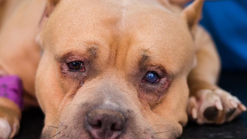
An eye ulcer, also medically termed corneal ulcer, or ulcerative keratitis in dogs, is a condition where the outer layer of the eye (the cornea) erodes, causing a divot or dent.
This divot can be superficial (on the surface), or it can affect the deeper layers of the eye. Eye ulcers lead to pain, redness of the eye, eye discharge, blinking frequently or holding the eye closed, and sometimes swelling. This is a common condition and is diagnosed most often in brachycephalic (flat-nosed) dog breeds but can easily affect any breed of dog.
Symptoms of Eye Ulcer in Dogs
Symptoms of eye ulcers in dogs range from mild to severe depending on the cause and severity of the erosion.
Symptoms in order of severity include:
-
Blinking the eye more often
-
Excessive tearing
-
Redness of the eye
-
Swelling of the eyelid or skin around the affected eye
-
Pawing at the eye or rubbing their face on the ground
-
Elevated third eyelid (third eyelid is located in the inside corner of the eye)
-
Holding the eye completely shut
-
Yellow/green/bloody eye discharge (in severe cases)
-
Hole (perforation) in the outer layer of the eye/rupture of the eye (in severe cases)
-
Blindness in affected eye (in severe cases)
-
Decreased appetite
-
Lethargy or hiding behavior
Causes of Eye Ulcer in Dogs
Anything that disrupts any part of the normal outer layer of the eye (cornea) can cause an ulcer, including if it affects the normal corneal structure, function, or physiology. Causes of eye ulcers in dogs are most commonly trauma, foreign body injury, or chemical burns.
These are some of the multiple causes of ulcerative keratitis in dogs:
-
Trauma: scratches from another animal; running into tree branches, plants, or bushes; self-inflicted trauma from rubbing their own face
-
Foreign body: sand, dirt, or other foreign material can enter the eye and get stuck behind the eyelids, causing repeated damage to the cornea
-
Chemical irritation/burns: shampoos, topical medications, or household cleaning products getting into the eye
-
Bacterial infections
-
Viral infections
-
Secondary to conformation/congenital issues of the eye: such as abnormal eyelash growth (distichia), eyelid masses or tumors, or entropion (rolling in of the lower eyelid)
-
Secondary to chronic dry eye: called keratoconjunctivitis sicca
-
Secondary to neurologic issues: that do not allow the eyes to blink appropriately
-
Secondary to certain endocrine diseases: such as diabetes mellitus, Cushing’s disease, and hypothyroidism
-
Secondary to inherited conditions: such as epithelial dystrophy, seen most commonly in Boxers, King Charles Cavaliers, Beagles, Cocker Spaniels, Afghan Hounds, and Alaskan Malamutes
How Veterinarians Diagnose Eye Ulcer in Dogs
Your veterinarian will want to perform some eye tests to find the cause of the ulceration and to decide on a course of action for treatment. The most important part of diagnosing and treating eye ulcers in dogs is to figure out if the erosion is simple or complicated.
A simple (uncomplicated) eye ulcer only involves the most superficial (surface) layer of the cornea and usually heals within 7-10 days without progression into the deeper corneal layers.
A complicated eye ulcer extends into the deeper layers of the cornea and can become infected or even start “melting” the deeper layers because of severe inflammation (swelling) and microorganism invasion.
There are three types of complicated ulcers:
Persistent corneal ulcers: This includes superficial ulcers with an elevated outer rim making it more difficult for the cornea to heal itself. They often heal with longer courses of therapy over several weeks to months. They sometimes require surgery to correct the problem if they worsen or do not heal with the right therapy.
Corneal foreign bodies: Objects can penetrate superficially or can penetrate the deeper layers of the cornea. The deeper the foreign body goes the faster and more intense inflammation occurs. Often these foreign bodies require careful removal by a veterinarian and aggressive therapy to heal. Surgery is sometimes necessary.
Stromal ulcers: These affect the deepest layer of the cornea, which is the wall between the outer layer of the eye and the fluid contained within the front chamber of the eye. They require aggressive medical and surgical management which may include melting ulcers (descemetoceles), and cat claw lacerations (cuts) or perforations (holes) to treat. These ulcers are often infected, severely inflamed, and painful.
Your vet will perform a thorough examination of the eye itself to assess for any foreign objects, obvious lacerations or abnormal eyelashes, and masses prior to any diagnostic testing.
Sometimes obvious lacerations or divots are noted in the eye with or without inflammation around the affected ulceration. Often, superficial erosions are not visible without special equipment.
Diagnostic testing that is usually performed by your veterinarian may include:
Fluorescein stain: A drop of this special yellow to green tinged dye is placed in your dog’s eye. The dye will adhere to the ulcerated tissue unless the ulcer is deep; then usually just the affected rim around the eroded area will uptake the dye. Small eye ulcers may require certain ophthalmic filter equipment to diagnose while other larger ulcers are often obvious when illuminated after fluorescein staining.
Schirmer tear testing: This is a simple, noninvasive test to measure tear production and to diagnose chronic dry eye in dogs. This is a common cause of corneal ulceration if no other obvious foreign material or objects are found to be causing the ulcer.
Intraocular pressures (tonometry): This is a simple, noninvasive test that helps to find the pressure behind the eye. If the corneal ulcer is deep or extensive, then this test may not be performed to avoid rupture of the eye itself. Often medical issues such as glaucoma or uveitis are diagnosed as a primary or secondary issue with deep or chronic corneal ulcers.
Bacterial culture: This involves taking a sample of cells from the cornea which are then allowed to incubate and checked for growth of bacterial cells. A culture helps to direct antibiotic therapy in melting, chronic, non-healing, and deep eye ulcers.
Cytology: This involves taking a small sample of cells from the cornea to assess under a microscope. A cytology can help direct the correct therapy in chronic, non-healing, or deep eye ulcers.
Treatment of Eye Ulcer in Dogs
Treatment of eye ulcers in dogs depends on the type of corneal ulcer and the severity of the ulcer itself. Therapy is divided into two types: medical and surgical therapies.
Medical Therapy for Eye Ulcer in Dogs
A therapy common for simple eye ulcers including broad-spectrum, topical antibiotics, and often topical pain medication. Sometimes systemic pain medication is added depending on the comfort level of the dog.
Contact lenses are sometimes used to protect the outer layer of the eye while uncomplicated ulcers are healing. Most of these simple ulcers heal with therapy within 3-7 days. It is important to continue watching even simple ulcers closely and follow up with your veterinarian for recommended recheck appointments to ensure that the ulcer is not getting deeper or becoming infected.
Complicated eye ulcers often require more potent topical antibiotics up to every 2-4 hours depending on the diagnosis. Oral antibiotics and pain medications are also often required for systemic therapy and comfort control. These types of ulcers are watched very closely with frequent veterinary exams to ensure that the ulcer does not worsen during therapy. These can progress to deep ulcers, which require surgical intervention.
Simple, superficial foreign objects can sometimes be easily removed from the cornea with ophthalmic irrigation (a sterile cleansing solution used to flush the eye), while deeper foreign bodies often need gentle removal under general anesthesia by a veterinarian. Remaining lacerations or ulcerations can be thoroughly treated after removal with aggressive medical therapy.
Indolent (non-healing) ulcers often require keratotomy or burr debridement, which uses a specific burring instrument (cutting tool) to remove the dead cells from the ulcer. It also stimulates blood vessels’ invasion into the ulcer for healing. Most of these ulcers heal about 2-3 weeks after this procedure.
Surgical Therapy for Eye Ulcer in Dogs
Surgical intervention is necessary when an eye ulcer is deep, complicated, melting, or is chronic and no longer responding to medical therapy.
Conjunctival (eye tissue) grafts are the most common surgery performed. This is where conjunctival tissue is transposed over the affected ulcer and is used to provide support and blood supply during healing. In some cases, a third eyelid flap and a temporary surgical closing of the eyelids (tarsorrhaphy) are used for ulcers caused by some congenital (present at birth) or neurologic (nervous system) issues. This acts as a bandage to protect the eye during healing.
Some chronic or deeper ulcers can cause secondary inflammation in the eye, or uveitis, which requires more aggressive anti-inflammatory therapy using systemic non-steroidal anti-inflammatory medications and additional systemic analgesics.
Elizabethan collars or pet cones are a necessity during healing from all eye ulcers. They eliminate the possibility of self-trauma caused by rubbing or pawing and protect your dog during healing. This collar must always remain on until it is deemed safe for removal by your veterinarian. Removing the collar too soon can lead to trauma and complicate current corneal ulcerations.
Recovery and Management of Eye Ulcer in Dogs
Most simple eye ulcers are rechecked by a veterinarian in 5-7 days to assess healing. Most of these ulcers heal during this time with the right therapy. Prognosis is usually excellent for full recovery.
Deeper or more complicated ulcers are checked more often, usually 1-2 days after diagnosis, to make certain that they are showing signs of improvement and not worsening. Sometimes, a pet may be hospitalized to perform frequent treatments every 2-4 hours. Medications are changed or extended based on bacterial culture and cytology results.
For melting, deep stromal, or complicated eye ulcers, referral to a veterinary ophthalmologist (eye doctor) is usually recommended for treatment and surgical intervention. Conjunctival grafts are usually left in place for 2-3 months. Complicated ulcers are monitored closely by a veterinarian for several weeks after surgery or therapy to assess for tear production, which may require medical management for short periods of time or permanently in some cases.
Any underlying medical conditions or congenital abnormalities must be diagnosed and correctly treated to avoid chronic eye ulcers or frequent development of corneal ulcers in the future.
Eye Ulcer in Dogs FAQs
What is the cornea?
The cornea is the clear cell membranous outer layer of the eye and is made up of three cell layers. The most outer layer is called the epithelium, the thick middle layer is the stroma, and the thinnest, innermost layer is the endothelium (otherwise known as Descemet’s membrane).
Can a corneal abrasion (scrape) progress to become a corneal ulcer?
Yes. Erosion of the outer layer of the eye, the corneal epithelium, is called a corneal abrasion or erosion. When this erosion gets deeper and affects the middle layer or inner layer of the eye, a corneal ulceration occurs. This usually happens from self-trauma (rubbing the face or eyes), bacterial infection, or chronic/progressive inflammation.
What is an indolent corneal ulcer?
Indolent corneal ulcers are ulcers affecting the superficial layer of the cornea that do not heal within 7 days. Often this type of ulcer will seem to be improving when therapy starts, but then it deteriorates with no obvious cause. Often, these ulcers require keratectomy (laser eye surgery) to remove the unhealthy layers and allow therapy and healing.
Featured Image: iStock.com/Todorean Gabriel