Narrowing of Vertebral Canal in Dogs

Lumbosacral Stenosis and Cauda Equina Syndrome in Dogs
A dog’s spine is composed of multiple bones with disks located in between adjacent bones called vertebrae. Seven cervical vertebrae are located in the neck (C1-C7), 13 thoracic vertebrae are present from the shoulder to the end of the ribs (T1-T13), seven lumbar vertebrae are present in the area starting from the end of the ribs to the pelvis (L1-L7), with the remaining vertebrae called sacral and coccygeal (tail) vertebrae.
Cauda equina syndrome involves the narrowing of the vertebral canal, resulting in compression of spinal nerve roots in the lumber and sacrum regions. Pressure to or damage of the nerves within the spinal canal in the junction area between the lumbar and sacral vertebrae (also called the cauda equine) due to narrowing of spinal canal can lead to this condition, also known as the cauda equina syndrome.
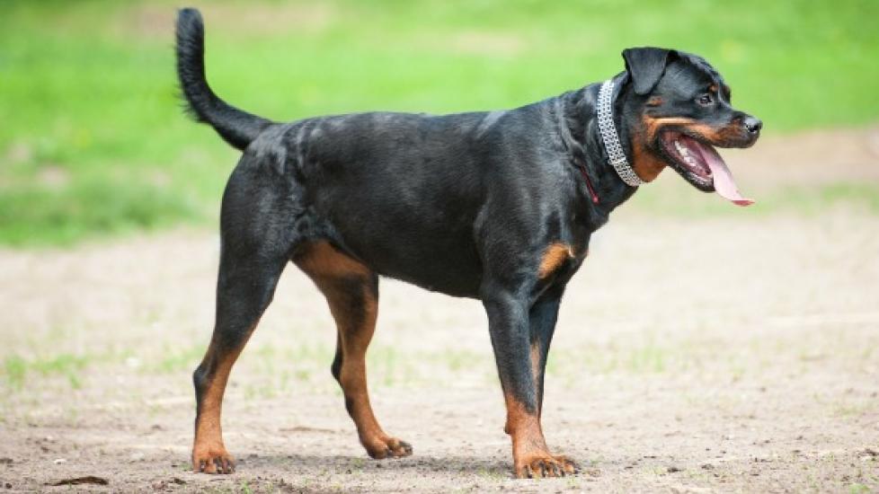
This syndrome is quite common in dogs. It can be congenital (present at birth) in small to medium-sized dogs, or an acquired condition (develops later in life), which is quite common in German shepherds, boxers, and rottweilers.
Symptoms and Types
- Lameness
- Pain in lumber and sacral regions
- Pelvic limb weakness and muscle wasting
- Weakness or paralysis of tail
- Abnormal tail carriage
- Urine and fecal incontinence (in some animals)
Causes
As stated earlier, cauda equina syndrome can either be a congenital or acquired condition, brought on by the instability of the lumbosacral junction or protrusion of disk between adjacent vertebrae.
Diagnosis
You will need to give a thorough history of your dog’s health, including the onset and nature of the symptoms, to your veterinarian. He or she will then perform a complete physical examination as well a biochemistry profile, urinalysis, and complete blood count -- the results of which are usually within normal range, unless some other concurrent disease is also present. Radiographic studies usually reveal valuable information for diagnosis. But for definitive diagnosis, your pet’s veterinarian will typically conduct Computed Tomography (CT-Scan) and Magnetic Resonance Imaging (MRI) testing.
Treatment
Dogs with urination problems are hospitalized for initial treatment (e.g., catheterization of bladder) until the patient regains control bladder function. Decompressing surgery is a treatment of choice and is often conducted to relieve the pressure of the nerve roots. If no treatment is conducted, the symptoms become severe due to progressive nature of this disease.
Even after surgery, however, some neurologic deficit may remain. Movements are restricted for at least four weeks after surgery. If surgery is not conducted, confinement and restricted leash walk is recommend along with pain control medications.
Living and Management
Avoid exercising your dog strenuously (jumping, running, etc.), as it may increase excessive pressure on the spine and cause symptoms to recur. Watch your dog for pain, lameness, urination and/or fecal elimination problems and notify your veterinarian immediately if you should notice any such untoward symptoms. Some diet modifications may also be recommended by your dog’s veterinarian to avoid obesity, which might also aggravate the condition.
Conform well to guidelines given by your dog’s veterinarian, especially directions regarding exercise, rest, and the diet of your dog.


