Gallbladder Obstruction in Dogs

Gallbladder Mucocele in Dogs
Gallbladder mucocele causes obstruction of the gallbladder's storage capacity due to the formation of a thick, mucoid bile mass inside the gallbladder, impairing its ability to function. The accumulated bile may extend the gallbladder, resulting in necrotizing cholecystitis – tissue death due to inflammation of the gallbladder.
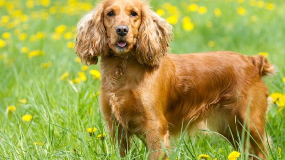
Gallbladder mucocele is common among middle-aged to older dogs, particularly Shetland sheepdogs, cocker spaniels and miniature schnauzers, and is not gender-specific.
Symptoms and Types
Gallbladder mucocele may be symptomatic or asymptomatic (without symptoms). The general symptoms are:
- Fever
- Vomiting
- Anorexia
- Dehydration
- Abdominal discomfort or pain
- Yellowish skin (jaundice)
- Polyuria/polydipsia (excessive urination/excessive thirst)
- Collapse – vasovagal or bile peritonitis (inflammation of the abdominal lining or dysfunction of the blood vessels)
Causes
- Lipid metabolism problems, particularly among Shetland sheepdogs and miniature schnauzers—this condition may be inherent in some dogs.
- Gallbladder dysmotility (lack of intra-organ movement)
- Cystic hypertrophy (abnormal enlargement) of the mucous-producing glands of the gallbladder, a common feature among older dogs—this condition may act as a trigger for gallbladder mucocele.
- High-fat diet, raised cholesterol or hyperthyroidism
- Typical or atypical adrenal hyperplasia – the abnormal multiplication of cells, and previous glucocorticoid therapy.
Diagnosis
The determining diagnosis of gallbladder mucocele will be based on the distinctive conditions that would cause abnormal functioning (dysmotility) of the gallbladder. Some of the possible factors responsible for bile blockage (stasis) are neoplasia (tumor growth), pancreatitis (inflammation of the pancreas), and choleliths (gallstones), amongst other observed causes.
Diagnosis is made through blood biochemistry, hematology, lab tests and imaging studies. The common observations are:
Biochemistry
- Analysis of liver enzymes, ALP, GGT, ALT and AST—high liver enzymes indicate illness. Sometimes, this may be the only sign of illness in dogs or it may manifest in the acute stage of the disease.
- Increased bilirubin
- Low albumin
- Electrolyte abnormalities with fluid and acid-base disturbances, which are due to excessive loss of fluids from vomiting or triggered by bile peritonitis.
- Pre-renal azotemia
Hematology/CBC
- Anemia
- Leukocyte imbalance
Lab tests
- High triglycerides
Imaging
- Radiography or ultrasound studies showing liver abnormalities, distended gallbladder and bile duct, gallbladder wall thickening, presence of gas in the liver, and loss of detail in the abdomen due to inflammation of the soft lining of the abdomen (peritonitis).
- The common diagnostic procedure is aspiration sampling of fluids drawn from biliary structures, or from the abdominal cavity, by use of laparotomy (incision into the abdominal cavity), liver biopsy, bacterial cultures and sensitivity tests, and cell examinations.
Treatment
Gallbladder mucocele treatment depends on the condition of the patient. Outpatients are generally put on anti-inflammatory and liver protecting agents like ursodeoxycholic acid and S-Adenosylmethionine (SAM-e). Inpatients are treated according to imaging and ultrasound results. Patients with higher lipids are restricted from fat-rich foods. If inflammation of the abdominal lining (bile peritonitis) is confirmed, abdominal cleansing (lavage) is recommended. All patients should be put on hydration therapy to correct fluid and electrolyte imbalances.
Other than broad-spectrum antimicrobials, depending on symptoms, the patients are put on anti-emetics, antacids, gastroprotectants, Vitamin K1 and antioxidant medications. After the treatment, all gallbladder mucocele patients must be periodically monitored with biochemistry, hematology and imaging studies to exclude/include various complications like cholangitis or cholangiohepatitis, bile peritonitis and EHBDO.


