Tumor Related to Vaccinations in Dogs

Vaccine-associated Sarcoma in Dogs
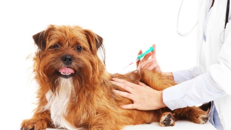
Most types of injectable vaccine and non-vaccine products have rarely been associated with sarcoma development in dogs, but some dogs may develop a site specific sarcoma following rabies vaccination. In fact, reports of a sarcoma (a cancerous mass arising from bone, cartilage, fat or muscle) developing at the site of vaccine injection sites in some animals have led to the suspicion of a link between the vaccine and a disposition in some animals to this type of reaction.
These tumors are characterized as highly invasive, rapidly growing, and malignant. Metastatic (spreading) rates are reported to be 22.5 to 24 percent. Often, the cancer spreads to the lungs, but it may spread to the regional lymph nodes and to the skin as well.
The cause for the sarcoma development is unknown, but it is believed that local inflammation must first occur for the malignant mass to follow. In addition, initial reports focused on vaccine adjuvants (assisting ingredients) containing aluminum as a potential cause of the sarcoma. However, the role of aluminum is unclear because not all adjuvants used in the vaccines that have been associated with sarcoma formation have contained aluminum.
Symptoms and Types
Lesions occur at the site of the vaccination, persisting and/or growing in size. In the advanced stages, the lesions will become fixed and occasionally ulcerated.
Causes
Vaccination with the rabies vaccine appears the be the underlying cause of this type of sarcoma. Moreover, the risk of developing the tumors may increase with the frequency and number of vaccinations given.
Diagnosis
You will need to give a thorough history of your dog's health, onset of symptoms, and possible incidents that might have precipitated this condition. Your veterinarian will order a blood chemical profile, a complete blood count, a urinalysis and an electrolyte panel.
To assess the spread of cancert, X-ray imaging of the chest and abdomen should be done. Computed tomography (CT) images with contrasting agents, meanwhile, is used because the agents enable to veterinarian to examine the area more readily.He or she can then record the location, shape, and size of all masses occurring at the injection sites.
Masses at vaccination sites that persist for longer than three months, are larger than two centimeters in diameter, or increase in size one month after the injection should be biopsied. Advanced lesions should also be biopsied prior to definitive treatment.
Treatment
An effective treatment protocol is difficult, but radiation therapy before or after definitive surgery will substantially enhance your dog's survival. Prior to surgery, a contrast CT scan should also be done, because it has been found to result in a substantially longer time until recurrence of the sarcoma. Chemotherapy, meanwhile, has not been found to enhance survival with this form of cancer.
Living and Management
Do not over-vaccinate your dog. Vaccinate for rabies and other diseases no more frequently than every three years, unless it has been specifically been advised by your veterinarian.


