Fibrosarcomas in Dogs


What Is a Fibrosarcoma in a Dog?
A fibrosarcoma is a type of cancer that forms in the connective tissue within the body. Connective tissue lines the entire body and helps to support and provide structure to all the muscles, ligaments, tendons, cartilage, nerves, bones, vessels, and fat. Fibrosarcoma tumors occur most often in the connective tissue beneath the skin.
Fibrosarcomas can also develop in the nose, mouth, and bones. If present in the limbs or mouth, they can invade the underlying bone, potentially leading to fractures.
Fibrosarcomas are a type of soft tissue sarcoma. They often grow slowly but can exhibit local aggressiveness by invading surrounding tissues. Only about 10% of fibrosarcomas will metastasize, meaning they will spread to other locations in the body such as the lymph nodes and lungs.
Fibrosarcomas are common in dogs, and large-breed dogs have a higher incidence of this type of cancer. Certain breeds, such as Irish Wolfhounds, Gordon Setters, Golden Retrievers, Brittany Spaniels, and Doberman Pinschers, may have a predisposition to develop fibrosarcomas.
Symptoms of Fibrosarcomas in Dogs
The clinical signs of fibrosarcomas depend on their location. Skin fibrosarcomas may manifest as a firm lump on the trunk of the body, while tumors in the mouth, nose, or bones may lead to drainage and pain in those areas. Dogs with fibrosarcomas commonly exhibit one or more of the following signs:
- Lump on the skin, often firm and sometimes ulcerated (bloody)
- Pain
- Lameness
- Nasal drainage
- Difficulty opening the mouth
- Poor appetite
- Halitosis (bad breath)
- Facial deformity
Causes of Fibrosarcomas in Dogs
The underlying causes of fibrosarcomas, like many other cancers, are not well understood. Cancer is generally believed to result from a combination of genetic and environmental factors. Chronic stimulation from implants, like bone plates from past surgeries, or foreign bodies can contribute to cancer development. Certain carcinogens, such as radiation exposure, are known to increase the risk of cancer. Genetics and hereditary factors play a significant role. In dogs, fibrosarcomas are more prevalent in large breeds and are more commonly diagnosed in older dogs, with the average age of 10 years.
Cancer refers to the unchecked division of cells. Normally, cell division occurs under the body’s guidance for specific purposes. In the case of cancer in dogs, cells divide independently, disregarding the normal regulatory mechanisms. Older animals are more susceptible to incorrect cell division, potentially having a higher number of mutated cells or a reduced efficiency in eliminating problematic cells.
How Veterinarians Diagnose Fibrosarcomas in Dogs
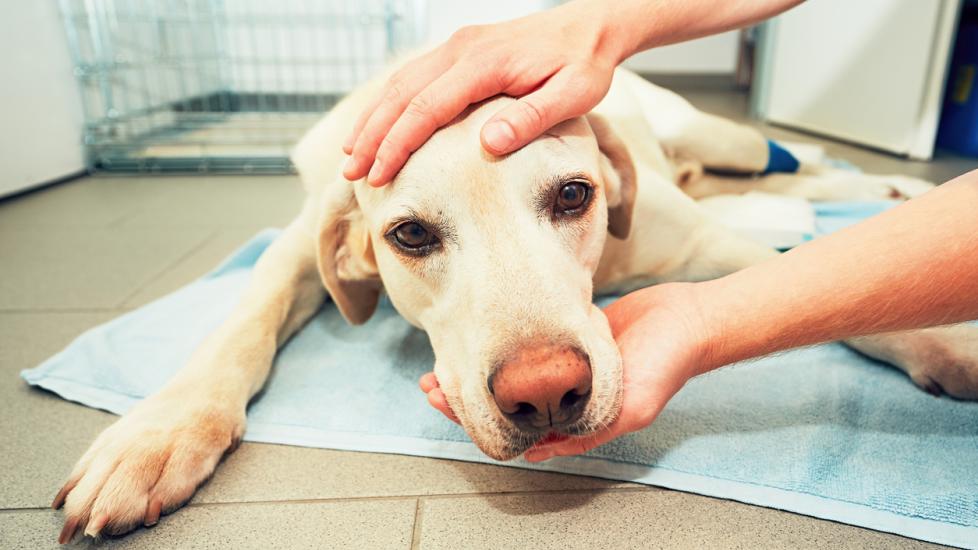
If your dog develops a new lump, it is important to have it examined by a veterinarian as soon as possible. It is not possible to determine whether a mass is cancerous or not by just looking at it. The best way to determine the precise cause of a lump is to examine the cells under a microscope.
The two most common tests to determine the cause of a lump are cytology and histopathology (biopsy). In cytology, your veterinarian will perform a fine needle aspirate (FNA). During this procedure, a needle is inserted into the lump to collect cells, which are then sprayed onto a slide and examined under a microscope.
Grading of Fibrosarcomas in Dogs
In addition to determining the type of cell present in a lump, examination also provides a grade for fibrosarcoma. Tumors are often graded and staged, with grading referring to the “what” and staging referring to the “where.” Grading involves determining the type of tumor and its likely behavior.
Fibrosarcomas can be categorized as Grade 1, 2, or 3, with Grade 1 tumors being the least aggressive and Grade 3 tumors being the most aggressive. More aggressive tumors require more intensive treatment. Once a fibrosarcoma is diagnosed and graded, additional tests are recommended for staging. Staging involves assessing the extent of the tumor’s progression and identifying the sites where the cancer has spread. Your veterinarian may recommend blood work, X-rays, an abdominal ultrasound, and possibly a CT scan to look for evidence that the cancer has spread. The lungs and lymph nodes are the most common sites of metastasis for fibrosarcomas.
Treatment of Fibrosarcomas in Dogs
The primary treatment of fibrosarcomas in dogs is typically surgery. Aggressive surgical intervention is often necessary to ensure complete removal of the cancer; however, it may not always be feasible to remove it all. For instance, a fibrosarcoma on a limb may require amputation of the entire leg to remove all the cancer cells. In cases where the tumor occurs in the nose, mouth, or body wall, complete tumor removal can be challenging or unviable.
If surgery is performed, the excised mass is sent for histopathology of not only the grade but also the margins. The term “margins” refers to whether the surgery removed all of the cancer. Ideally, normal tissue surrounding the mass is removed to ensure that no cancer cells have infiltrated the surrounding tissue, minimizing the risk that the cancer can recur.
Sometimes surgery is not an option, such as when the tumor is excessively large and it would be impossible to close the skin if it was removed. This is more often the case when fibrosarcomas occur in locations such as the mouth. In these cases, radiation therapy may be recommended. Radiation can also be administered prior to surgery to shrink the tumor size. A pup’s age and other underlying health conditions are also reasons why surgery may not be recommended.
Additionally, oral medications may be prescribed to alleviate pain, reduce swelling, or treat secondary infections, especially if the mass is ulcerated. While fibrosarcomas generally have a limited response to chemotherapy drugs, there are instances where pain medications may be recommended as an alternative when surgery isn’t feasible. Common medications include:
Recovery and Management of Fibrosarcomas in Dogs
After surgery, it is important to keep your dog’s activity level restricted to avoid excessive stress on the incision. Usually, the post-op recovery time is two weeks. A recovery cone will likely be sent home to prevent your dog from licking or chewing the incision or rubbing the surgery site. Be sure to keep the incision clean and dry and let your veterinarian know if there is any unusual drainage or if your dog becomes lethargic or will not eat.
If surgery is not possible, the management approach focuses more on keeping your dog comfortable. This may involve the administration of pain medications or antibiotics if secondary infections develop. Probiotics may also be beneficial for maintaining a balanced intestinal flora in dogs receiving pain meds or antibiotics, whether it is during the post-operative period or for supportive care.
The prognosis for fibrosarcoma can vary dramatically, depending on factors such as the tumor’s location, its aggressiveness, and the extent of spread at the time of diagnosis. Although fibrosarcomas tend to invade nearby tissue, they are less prone to metastasize compared to many other malignant cancers. This characteristic can result in longer survival times and a more favorable prognosis with appropriate treatment.
Fibrosarcomas in Dogs FAQs
How long can a dog live with fibrosarcoma?
Survival times vary depending on where the fibrosarcoma is located, how aggressive that particular tumor is, and how far it has spread when it is diagnosed. A dog can live two to four more years with lower-grade tumors with complete excision, compared to less than a year with higher-grade tumors and incomplete excision.
Is fibrosarcoma curable in dogs?
Fibrosarcomas can be curable in dogs in cases where they are low-grade and completely removed. While a complete cure is less common with this cancer type, it is a possibility.
How fast do sarcomas grow in dogs?
Fibrosarcomas in dogs are generally slow-growing. However, they are highly invasive and tend to infiltrate nearby tissues.
Featured Image: iStock.com/Chalabala
References
Bostock, D., Dye, M. National Library of Medicine. Prognosis after surgical excision of canine fibrous connective tissue sarcomas. 1980.
Dobson, J. National Library of Medicine. Breed-Predispositions to Cancer in Pedigree Dogs. 2013.
Fox-Alvarez, S. Clinician’s Brief. Tumor Grading and Staging in Dogs. 2022.