Mast Cell Tumors in Cats


What Are Mast Cell Tumors in Cats?
Mast cells are a type of white blood cell present throughout the body, including the skin, respiratory tract, and digestive tract. These cells contain histamine, a chemical compound that plays an important role in the immune system’s defense against perceived allergens.
When a mast cell is exposed to an allergen, it goes through a process called degranulation and releases histamine, which—to remove the allergen from the body—causes allergy symptoms such as sneezing, itching, and a runny nose and/or eyes. When histamine is released in excessive amounts, however, it can cause anaphylaxis—a life-threatening allergic reaction.
A mast cell tumor (MCT), is a type of tumor that arises from the rapid replication and division of mast cells in the tissue. Mast cell tumors can grow slowly over time, or develop very rapidly, seemingly overnight.
Mast cell tumors in cats are found on the skin (cutaneous MCTs) in most cases, typically appearing on the head and neck—but can appear anywhere on the body. These are the second most common type of skin tumor to develop in cats. Mast cell tumors can also develop on the spleen (visceral or splenic MCTs) and within the intestinal tract (intestinal MCTs), typically the small intestines.
Are Mast Cell Tumors Cancerous?
Tumors can be benign or malignant:
-
A benign tumor or growth is non-cancerous and does not invade surrounding tissues, or spread to other parts of the body. These lesions are typically slow growing and have an favorable prognosis, overall.
-
A malignant tumor or growth is cancerous and comprised of abnormal cells that tend to divide rapidly. These cells invade the surrounding tissues readily. Its spread to other areas of the body can also occur via the bloodstream or lymphatic system. Malignant tumors tend to be aggressive and generally have a poor long-term prognosis.
Mast cell tumors in cats can be benign or malignant depending on the characteristics of the cells present and where they are located. For example, mast cell tumors on the skin tend to be benign, while mast cell tumors affecting the internal organs are more likely to be malignant.
Symptoms of Mast Cell Tumors in Cats
Clinical signs and symptoms can vary depending on where the mast cell tumor is located and how aggressive (if malignant) the tumor is. Mast cell tumors on the spleen, or within the intestinal tract, will be more likely to cause systemic (whole-body) signs:
-
Cutaneous:
-
Hard, hairless, flattened bumps (plaques) located commonly on the head and neck, but can develop anywhere on the body
-
Small lumps (nodules) that feel very firm within the skin
-
Excessive itchiness and associated hair loss
-
Ulceration (an open sore)
-
-
Splenic or visceral:
-
Vomiting
-
Weight loss
-
Poor appetite
-
-
Intestinal:
-
Vomiting and diarrhea
-
Fresh blood in the stool
-
Black or tarry stool, indicating digested blood
-
Causes of Mast Cell Tumors in Cats
While the exact cause of mast cell tumors is not fully known, both genetics and environmental factors are suspected to play a role. For example, Siamese cats tend to be prone to the development of cutaneous mast cell tumors.
In dogs, there is a mutation present in a specific gene, referred to as KIT, that’s associated with the development of mast cell tumors. The KIT protein plays a role in cell replication and division. A mutation in the KIT has also been detected in cats with mast cell tumors.
How Veterinarians Diagnose Mast Cell Tumors in Cats
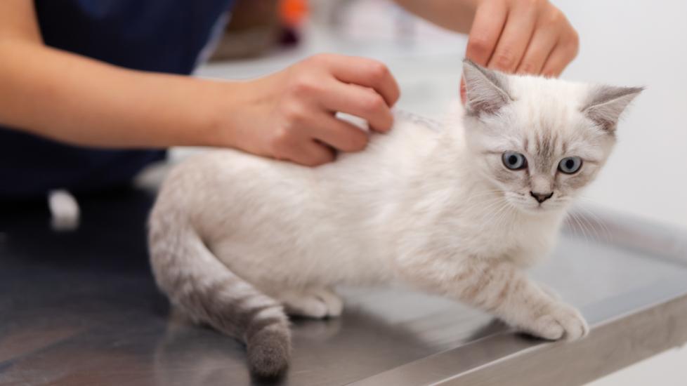
Mast cell tumors can be diagnosed by a procedure called a fine needle aspiration (FNA). A sterile needle is inserted into the tumor and a sample of the cell is retrieved using a syringe. Your veterinarian, or a veterinary pathologist, will then examine the cells under a microscope called cytology. Mast cells have a distinct appearance and abnormalities are typically easily diagnosed via cytology.
In some cases, your veterinarian may recommend a biopsy of the tumor. A sample of tissue from the tumor, or the entire tumor, can be removed and submitted for histopathology review by a veterinary pathologist. The histopathology report will provide your veterinarian with a more detailed description of the cells present within the tumor. This will help determine the type of tumor, how aggressive it is, and if clean surgical margins are possible. Clean surgical margins means that no cancer cells are still present on the outer edges of the tissue when surgically removed. This is the main goal when excising a tumor to prevent recurrence of growth.
If systemic illness is present or a malignant type of mast cell tumor is suspected or confirmed, your veterinarian may also recommend additional diagnostics, such as lab work (i.e., bloodwork, urinalysis) and imaging of the chest and abdomen. This will help determine the stage of the disease process, plan the most appropriate treatment protocol, and gauge your pet’s overall prognosis.
Treatment of Mast Cell Tumors in Cats
The recommended treatment protocol will depend on where the MCT is located and how invasive the tumor is. The choice of treatment for cutaneous mast cell tumors is surgical removal.
Complete surgical removal of splenic and intestinal mast cell tumors may not be obtainable and cancerous cells can be left behind. Sometimes cutaneous mast cell tumors in cats can spread to other areas of the body, though unusual. Alternative treatment options may be recommended in these cases, such as chemotherapy or radiation. This will likely require a referral to a veterinary oncologist.
When pierced or ruptured (as by aspiration or surgery), all mast cell tumors can cause systemic symptoms in your pet due to the release of histamine, and therefore your veterinarian will likely prescribe medications to avoid any negative side effects during treatment. Antihistamines, antacids, anti-nausea, and pain medications are common examples.
Recovery and Management of Mast Cell Tumors in Cats
Most cats with cutaneous mast cell tumors will go on to live healthy, normal lives. Recurrence of cutaneous mast cell tumors can happen, but it is rare and generally occurs when inadequate (non-clean) surgical margins were obtained.
Cats that develop splenic or intestinal mast cell tumors typically have a poor long-term prognosis since these types tend to be malignant and spread to other areas of the body more readily. Daily medications to help manage systemic illness, and improve quality of life, may also be recommended in these cases.
If your pet is diagnosed with a MCT, surgery will likely be recommended as a treatment option, and it is important to follow your veterinarian’s instructions once your pet is discharged. An E-collar (recovery cone) will likely be needed to prevent any self-trauma or injury to the surgical site while your pet is healing post-operatively.
It is also important to keep the surgical site clean and dry, and to limit your pet’s activity for at least 10-14 days to prevent dehiscence (opening of the wound). If you have any concerns while your pet is recovering at home, promptly contact your veterinary clinic.
Featured Image: iStock/ardaayderman